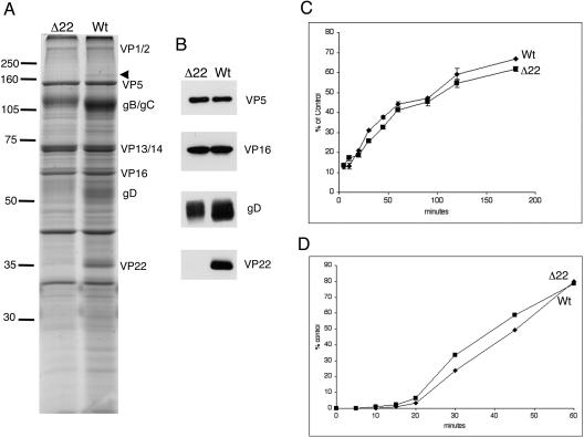
FIG. 3.
Characterization of HSV-1 virions assembled in the absence of VP22. (A) Equivalent amounts of purified extracellular WT and Δ22 virions were analyzed by SDS-PAGE and stained with Coomassie blue. Major virion proteins are labeled. The arrowhead denotes a species around 175 kDa that is absent from Δ22 virions. Numbers on the left indicate molecular mass in kilodaltons. (B) The same samples shown in panel A were analyzed by Western blotting with antibodies against the indicated virion proteins. (C) Monolayers of Vero cells were infected at 4°C with approximately 300 PFU of virus stock. At the indicated times, the cells were washed and transferred to 37°C, and plaques were allowed to develop. (D) Monolayers of Vero cells were infected with approximately 300 PFU of virus stock and incubated at 4°C for 2 h. Cells were transferred to 37°C and washed with citrate buffer (pH 3) at the indicated times, and plaques were allowed to develop.

