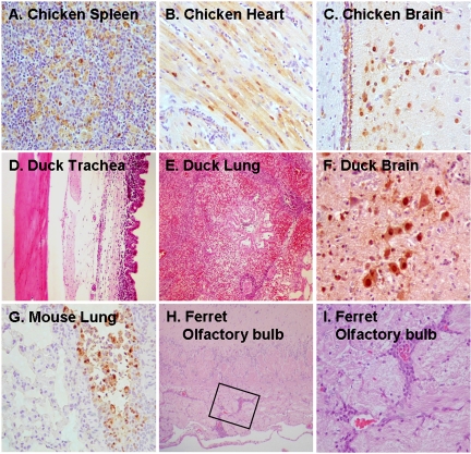
FIG. 2.
Representative histologic and immunohistologic findings on animals infected with HK213Egg virus. Chickens: detection of viral antigens (brown) in macrophages in the spleen (A), cardiac myocytes (B), and brain tissue (C) on day 2 p.i. Ducks: severe infiltration of mononuclear cells into the lamina propria mucosae of the trachea (D) and focal pneumonia with prominent infiltration of mononuclear cells (E) in asymptomatic animals and brain lesions with prominent viral antigen expression in the dead duckling (F). Mice: antigen-positive cells along the bronchial epithelium on day 3 p.i. (G). Ferrets: perivascular infiltrations of mononuclear cells in the olfactory bulb (H). The boxed area in panel H is shown at a higher magnification in panel I.

