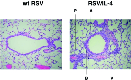
FIG. 2.
Histopathology in the lungs of mice after primary infection with wt RSV or RSV/IL-4. Mice were infected with RSV/IL-4 or wt RSV and sacrificed on day 4. The lungs were removed, fixed, cut along the midcoronal plane, and stained with hematoxylin and eosin. In animals infected with wt RSV (left), only a minimal amount of lymphocytic infiltration around the airways and blood vessels was observed, and there was no inflammation in the alveoli and alveolar walls. In contrast, in animals infected with RSV/IL-4 (right), a cellular infiltrate consisting primarily of lymphocytes was readily apparent around the airways (peribronchiolitis, indicated by B) and blood vessels (perivasculitis, V). A cellular infiltrate consisting primarily of lymphocytes and macrophages was also observed in the alveoli (alveolitis, A) and alveolar walls (interstitial pneumonitis, P). Magnification, ×100.

