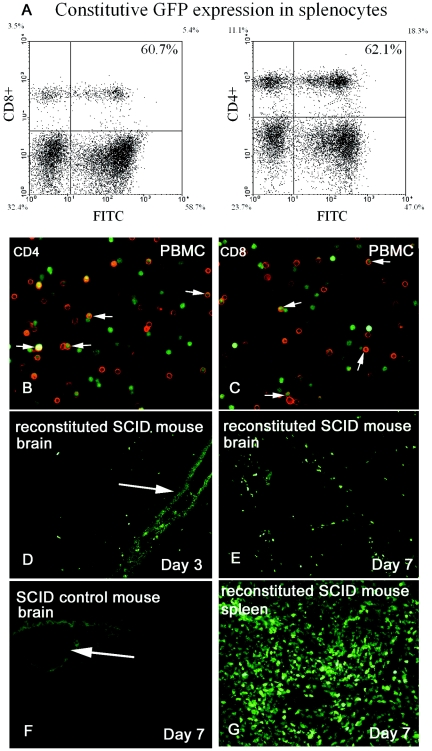
FIG. 3.
In vivo tracking of repopulated lymphocytes. Lymphocytes constitutively expressing GFP were used to reconstitute SCID mice infected with wild-type MCMV. (A) FACS analysis of constitutive GFP expression in splenocytes from transgenic Swiss Webster mice. Peripheral blood CD4+ (B) and CD8+ (C) T lymphocytes (red) constitutively expressing GFP; arrows, double-stained cells. (D to F) Cryosections of SCID mouse brain following intracranial infection of MCMV; arrows highlight the injection track. Activate lymphocytes (green) translocate across the blood-brain barrier within 3 days of systemic reconstitution and are seen in proximity to CMV infection. (E and F) SCID mouse brain 3 days (E) and 7 days (F) following reconstitution with CMV-immune splenocytes. F) SCID mouse reconstituted with medium; arrows highlight the injection tract. (G) Lymphocyte (green)-repopulated SCID mouse spleen 7 days following reconsititution.

