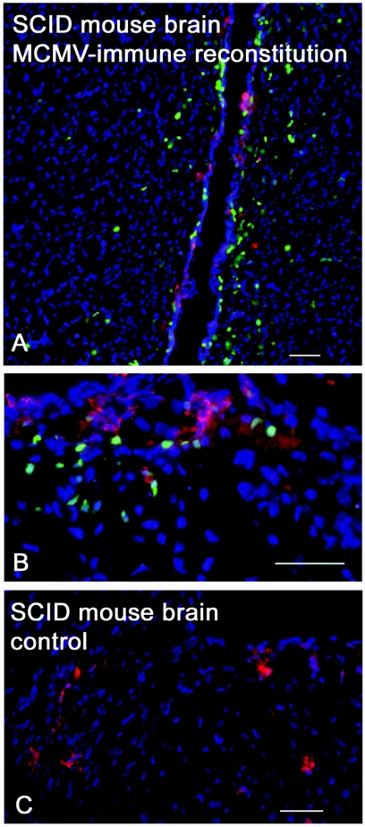
FIG. 4.
Activated lymphocytes invade brain and target antigen. Immunohistochemistry of stained cryosections of SCID mouse brain 8 days following MCMV challenge and 7 days following adoptive transfer of unsorted MCMV-immune splenocytes expressing GFP. (A) Periventricular migrating lymphocytes. (B) Foci of lymphocytes targeting MCMV infection in the cortex. (C) Control SCID mouse with active MCMV infection. Green, activated lymphocytes; red, MCMV antigen; blue, 4′,6′-diamidino-2-phenylindole stain of cellular nuclei. Bar, 50 μm.

