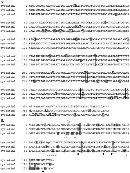
FIG. 1.
Nucleotide and amino acid sequence alignment of the CcBV cystatins. (A) Nucleotide sequence alignment of the three CcBV cystatin ORFs. Differences in nucleotide sequences are boxed. (B) Amino acid sequence alignment of the three CcBV preprotein cystatins. The potential cleavage site for cystatin 2 and cystatin 3 (same site) and cystatin 1 are indicated by white triangles. The three active sites which are the hallmarks of cystatins are indicated by stars: G (residue 28), Qx(V)xG (starting residue 72), and W (residue 119). The four cysteine residues potentially forming two disulfide bonds are indicated by black diamonds. Differing amino acids are boxed (same family, light grey boxing; different family, dark grey boxing).

