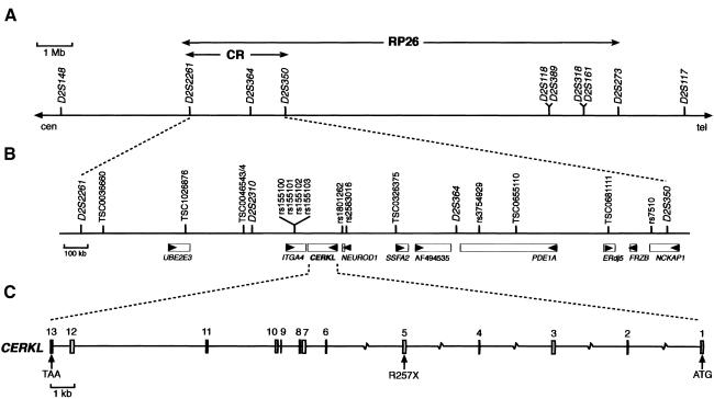
Figure 1.
The RP26 locus and the CERKL gene and protein. A, RP26 physical map of the 12.5-Mb cosegregation interval between markers D2S2261 and D2S273. B, Localization of CERKL within the candidate region (“CR”) defined by homozygosity mapping. Genes (rectangles) and the transcription direction (arrowheads) are indicated. C, CERKL exon/intron structure. The location of the R275X null mutation in exon 5 is shown.

