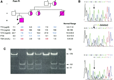
Figure 2.
Family Fi. A, Pedigree and thyroid function tests. For details and abbreviations, see the legend to figure 1. B, Electropherograms showing the WT reverse sequence from part of exon 3 of the MCT8 gene (upper tracing), the location of the nucleotide deletion in the propositus (middle tracing), and the sequence of the heterozygous mother (lower tracing). C, Results of genotyping of all family members for the mutation shown in B. The 178-bp product amplified from the mutant allele is digested into two fragments of 101 bp and 77 bp by HhaI, whereas the WT allele remains intact. Complete digestion is observed in the two hemizygous affected males (II4 and III1). The heterozygous females (I2, II2, and II3) show three bands: the 179-bp fragment representing the WT allele and the two smaller fragments representing the digested mutant allele.

