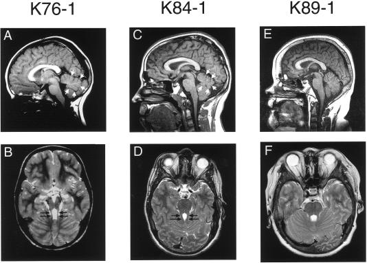
Figure 1.
Midline sagittal T1 images (A, C, and E) and associated axial T2 images through the level of the superior cerebellar peduncles (SCPs) (B, D, and F) in subjects indicated above each column. Note the superiorly positioned fourth ventricle with moderate inferior vermis hypoplasia (small white arrows) on the sagittal images of subjects K76-1 (A) and K84-1 (C), compared with the normal fourth ventricle with mild inferior vermis deficiency in subject K89-1 (E). Elongated SCPs are demonstrated on axial images (B and D, black arrows), with associated mild MTS, compared with normal SCPs (F).

