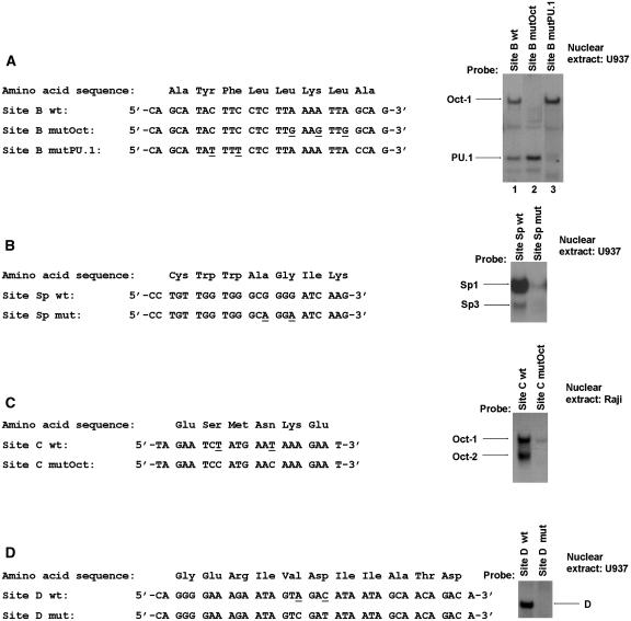
Figure 9.
Mutagenesis of the HS7 binding sites. Left panels: The wild-type and mutated oligonucleotides corresponding to the HS7 site B (A), Sp site (B), site C (C) and site D (D) are shown. The amino acids encoded by these oligonucleotide sequences are indicated. Bases that are changed in the mutated versions of the HS7 binding sites relative to the wild-type version are underlined. Right panels: Probes (indicated at the top of each lane) were incubated with 15 μg of nuclear extracts from U937 (A, B and D) or Raji (C) cells. The figure shows only the specific retarded bands of interest. The retarded complexes corresponding to Oct-1, Oct-2, PU.1, Sp1, Sp3 and D are indicated by arrows.

