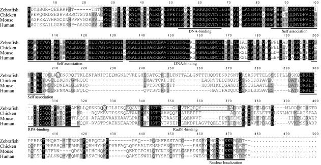
Figure 1.
Multiple alignment of Rad52 from zebrafish and other species. Multiple alignments were made using the DNASIS software (Hitachi Software Engineering). The longer splice isoform of zebrafish Rad52 is shown. Amino acid residues conserved in three or four species are highlighted in gray or black, respectively. Known domains/regions are underlined. DNA-binding—regions that are involved in single-strand DNA binding. The amino acid sequence that is deleted in the short isoform is indicated by an open gray box. Amino acids Q204 and Q317 are indicated by open gray ovals.

