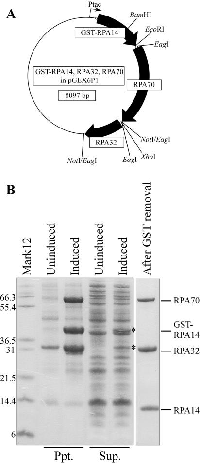
Figure 3.
Expression and purification of RPA proteins. (A) Schematic drawing of tricistronic expression construct as described in Materials and Methods. Ribosome binding sites inserted upstream of each open reading frame are depicted as small open circles. Restriction enzyme sites that were used for construction are shown. (B) SDS–PAGE analysis of RPA proteins from induced and uninduced cultures fractionated into precipitate (Ppt.) and supernatant (Sup.). Most RPA proteins were found in the precipitate, with small fractions of GST-RPA14 and RPA32 also found in the supernatant (asterisks). The final preparation of RPA after refolding and GST removal is shown in the right lane. Mark12; molecular weight marker.

