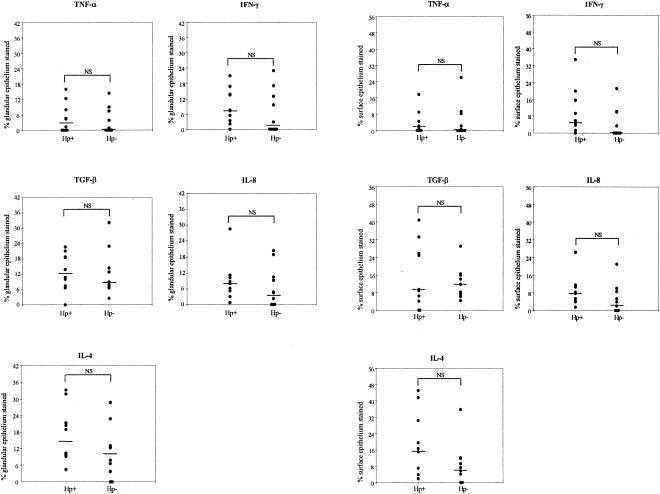
FIG. 3.
Epithelial cytokine staining in H. pylori-infected (Hp+) and uninfected (Hp-) subjects. Staining of both superficial epithelium (left panels) and epithelial cells from glands (right panels) were expressed as a percentage of the total surface and glandular epithelial area in the section, respectively. Each circle represents a biopsy specimen from one individual. Bars represent median values. NS, not significant (Mann-Whitney test).

