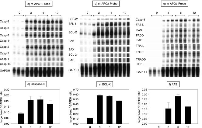
FIG. 2.
Increased thymocyte expression of apoptotic genes in response to LPS. Wild-type mice received an i.p. injection of 50 μg of LPS, and their thymuses were harvested at 3, 6, and 12 h later. The 0-h time point presents data from animals injected with a similar volume of PBS. RNA was extracted from thymocytes, and the genes involved in apoptosis were detected by RPAs. Here we show the autoradiograph of three RPA gels done with commercially available probe sets. The bands were analyzed with an imaging program. Below each gel is a bar graph that corresponds to the analysis of a chosen gene (d to e, respectively). The genes were chosen based on their relevance (caspase-3 and FAS) or level of expression (BCL-X). Data are expressed as the ratio of the mean density of the target band over the mean density of the housekeeping gene GAPDH. As a rule, LPS caused a marked and significant increase in the level of expression of the mRNAs of all apoptotic genes tested.

