
Figure 7.
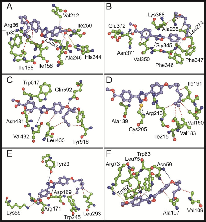
Molecular interactions of the title compound with different microbial targets. Molecular interaction of the title compound with (A) E. coli FabH (PDB id: 1HNJ), (B) S. aureus FmtA (PDB id: 5ZH8), (C) S. mutans glucosyltransferase (PDB id: 3AIE), (D) S. mutans sortase A (PDB id: 4TQX), (E) C. albicans Als3 adhesion protein (PDB id: 4LEB), and (F) T. brucei methionyl tRNA synthetase (PDB id: 4MW2) are shown in ball-and-stick representation. The carbon is shown in lavender for the title compound and green for amino acids. The red- and blue-colored atoms in all molecules indicate oxygen and nitrogen atoms, respectively. H-bonds are represented by blue dotted lines, and red dotted lines represent hydrophobic interactions. π–π interactions and slat bridge interactions are shown in black and green dotted lines, respectively.