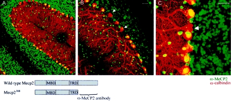
Figure 2.
Determination of XCI patterns in the cerebellum of Mecp2308/X females by confocal laser scanning microscopy. Costaining with anti-calbindin antibody (red) and an antibody against the C-terminus of MeCP2 (green) (see lower diagram for antigen localization) allows us to label all PCs (calbindin) and identify neurons that express the wild-type Mecp2 allele (cells positive for calbindin and Mecp2 have yellow nuclei). Note that the majority of PCs are immunoreactive for the MeCP2 antibody. Arrows mark the few cells expressing mutant Mecp2 (nonimmunoreactive for α-MeCP2). Each panel is a representative example of immunofluorescence, visualized at increasingly higher magnifications. Scale bar = 20 μm.

