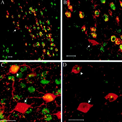
Figure 4.
XCI patterns in midbrain and cerebral cortex of Mecp2308/X female mice. A and B, Double immunofluorescence for the detection of wild-type MeCP2 (green) and TH (red) was used to determine the XCI pattern in catecholaminergic neurons in the midbrain. Arrows denote cells that have the X chromosome bearing the mutant Mecp2 as the active one. C, Visualization of coimmunolabeling of Mecp2 (green) and parvalbumin (red) in the cerebral cortex by confocal microscopy. D, Same as in panel C, but a single focal section is shown, to demonstrate the definitive identification of cells expressing the wild-type (arrowhead) versus the mutant (arrow) Mecp2 allele. Scale bar = 20 μm.

