Figure 7.
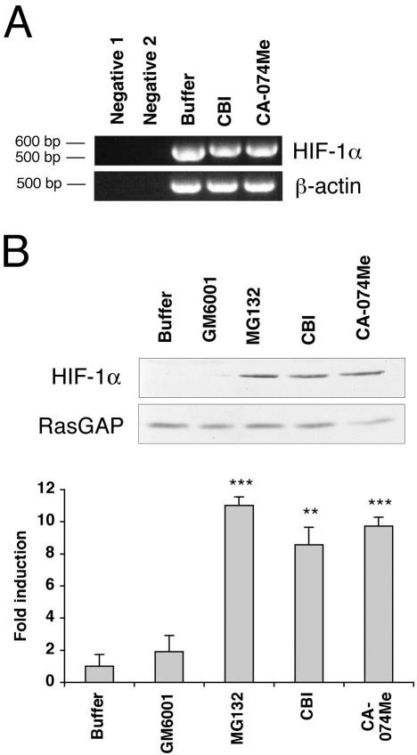
The cathepsin B inhibitors stabilize HIF-1α. (A) BRECs were plated in a collagen sandwich gel, and then medium containing either buffer or cathepsin B inhibitors (40 μM CBI or 1 μM CA-074Me) was added. After 1 d, total RNA was isolated and subjected to RT-PCR using primers specific for bovine HIF-1α. Bovine β-actin was amplified simultaneously in a separate set of tubes under the same conditions. (B) The BRECs were plated in a collagen sandwich gel, and then medium containing either buffer or cathepsin B inhibitors (40 μM CBI or 1 μM CA-074Me) or a proteasome inhibitor (10 nM MG132) or an MMP inhibitor (1 nM GM6001) was added. After 8 h in culture at 37°C, the cells were harvested. Total cell lysates were made and subjected to Western blot analysis using an anti-HIF-1α antibody. The blot was reprobed with an anti-RasGAP antibody to determine the level of protein loaded in each lane. The bar graph below is a fold increase in an intensity of HIF-1α band compared with buffer-treated control. Error bars, SD of three independent experiments. For the comparison of inhibitor-versus buffer-treated samples: ** p < 0.01; *** p < 0.001.

