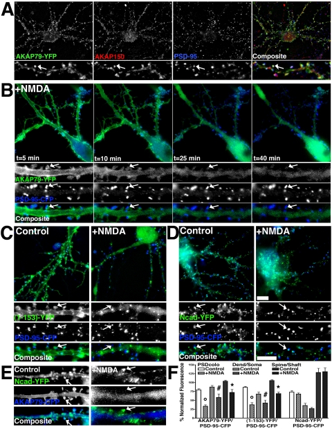
Figure 10.
NMDA receptor regulation of AKAP79 postsynaptic localization with PSD-95 and Ncad imaged in living neurons. (A) AKAP79-YFP (green) colocalizes (arrow, white) on dendritic spines with endogenous rat AKAP150 (red) and PSD-95 (blue) in hippocampal neurons. (B) Time-lapse imaging in living hippocampal neurons of AKAP79-YFP (green) redistribution (arrows) away from PSD-95-CFP (blue) on dendritic spines in response to NMDA (50 μM, 33°C, 5–40 min). (C) Postsynaptic localization of AKAP79(1-153)-YFP (green) with PSD-95-CFP (blue) on dendritic spines (arrows) seen in control conditions is lost in response to NMDA. (D) Postsynaptic localization of Ncad-YFP (green) with PSD-95-CFP on dendritic spines (arrows) is unchanged in response to NMDA. (E) AKAP79-CFP (blue) redistributes away from Ncad-YFP (green) on dendritic spines in response to NMDA (arrows). (C–E) Post-NMDA 30–40 min; smaller panels in A–E show magnifications of dendrites, arrows mark spines where CFP/YFP colocalization is seen in control neurons but is not seen after NMDA treatment). (F) Quantitation (n = 10 cells) showing significant decreases in NMDA-treated relative to untreated controls for AKAP79-YFP and (1-153)-YFP but not Ncad-YFP localization to dendrites relative to the soma (dend/soma ratio), dendritic spines relative to the cytoplasm of dendrites shafts (spine/shaft ratio) and colocalization with PSD-95-CFP (PS-Dcolo). Bars, 10 μm.

