Figure 3.
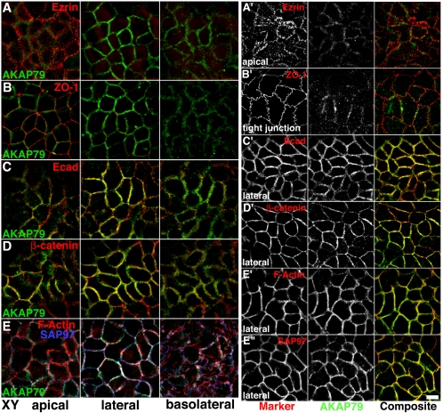
Polarized localization of endogenous AKAP79 with E-cadherin at the lateral membrane adherens junctions of Caco-2 epithelial cells. Lack of colocalization of AKAP79 (green) with apical membrane ezrin (red) (A and A′) and tight junction ZO-1 (red) (B and B′). Lateral membrane colocalization (yellow) of AKAP79 (green) with Ecad (red) (C and C′) and β-cat (red) (C and C′). (E, E′, and E″) Lateral membrane colocalization (white or yellow) of AKAP79 (green) with cortical F-actin (red, E and E′) and SAP97 (E, blue; E″, red). (A–E) xy, composite images through apical, lateral, and basolateral sections, as labeled. (A′–E″) Individual channels and composite images for a single xy section through apical, lateral, and basolateral membranes as labeled. Bar, 10 μm.

