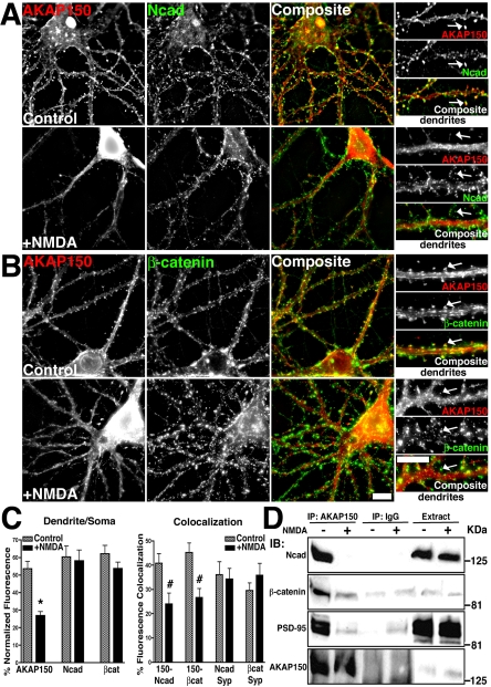
Figure 9.
Regulation of AKAP79/150–cadherin complexes by NMDA receptor activation. (A and B) Loss of AKAP150 (red) dendritic spine colocalization (yellow) with Ncad (green) (A) and β-cat (green) (B) in response to NMDA (50 μM, 37°C, 10 min) in hippocampal neurons. Smaller panels show magnification of dendrites, arrows point to colocalized signal in control spines and loss of spine colocalization after NMDA treatment. (C) Quantitation (n = 10 cells) showing a significant decrease in AKAP150, but not Ncad or β-cat, localization to dendrites relative to the soma (dendrite/soma ratio, left-hand graph) and a significant loss of AKAP150 punctate colocalization with Ncad and β-cat (colocalization, left-hand graph) with NMDA. Colocalization of Ncad and β-cat with the presynaptic marker Syp was unchanged by NMDA (n = 7 cells). (D) Disruption of AKAP79/150 association with cadherins, catenins, and PSD-95 MAGUKs in response to NMDA. Approximately 7 mg of Triton-soluble extracts from control untreated (–) and NMDA treated (+) neurons were immunoprecipitated (IP:) with either 5 μg of anti-AKAP150 or nonimmune IgG as indicated. IPs and cell extracts were probed (IB:) as indicated. Bars, 10 μm.

