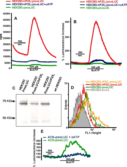
Figure 3.
ATP release occurs through the P2X7 receptor. (A and B) HEK293 cells cotransfected with hP2X7 and pmeLUC (HEK293-hP2X7/pmeLUC) were placed in the luminometer chamber and perfused with a BzATP-containing solution, with (blue trace) or without (red trace) prior treatment with 300 μM oATP for 2 h. HEK293-pmeLUC (green trace) are shown as a control. Luminescence increase is expressed as cps in A and percent increase over basal in B. In C, total pmeLUC protein expressed in HEK293-pmeLUC, HEK293-hP2X7/pmeLUC, and HEK293-rP2X7/pmeLUC is determined by Western blotting. wtHEK293 are shown as a control. In D, plasma membrane-expressed pmeLUC is measured by FACS analysis in the three cell populations. ATP release from human neuroblastoma ACN cells incubated in the absence (green trace) or presence (blue trace) of 300 μM oATP is shown in E.

