Figure 7.
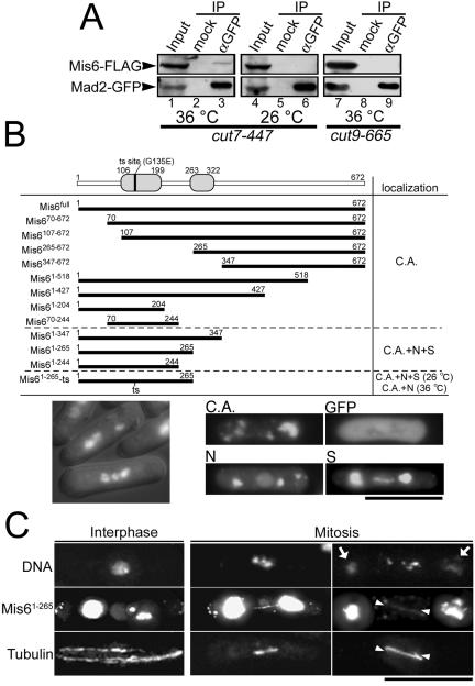
Mis6 physically interacts with Mad2. (A) Immunoprecipitation experiments were performed using crude extract from cut7-477 or cut9-665 mutant cells harboring integrated mis6+-FLAG and mad2+-GFP genes. The cut7 mutant cells were cultured at either 36°C for 3 h (restrictive condition, lanes 1–3) or 26°C (permissive condition, lanes 4–6). The cut9 mutant cells were cultured at 36°C for 3.5 h (restrictive condition, lanes 7–9). The anti-GFP mAb (Roche Diagnostics) was used for the precipitation of GFP-tagged Mad2 (lanes 3, 6, and 9). In the mock experiment, the buffer was added instead of the antibody (lanes 2, 5, and 8). Twenty percent of the inputs were loaded on lanes 1, 4, and 7. (B) A series of truncated Mis6 were fused with GFP and ectopically expressed under the nmt1-1 promoter, and their subcellular localization was determined. C.A., N, and S stand for cytoplasmic aggregate formation, nuclear localization, and spindle localization, respectively. N-terminal domains that are highly conserved among fission yeast, chicken, and human were indicated by gray boxes in the schematic drawing of Mis6 protein. The mutation site of Mis6-302 temperature-sensitive protein also was indicated by a vertical bar. Representative images of the truncated Mis6 constructs forming cytoplasmic aggregates, localizing in the nuclei or localizing along the mitotic spindle were shown in right bottom panels. An image of ectopically expressed GFP alone, which dispersed throughout the cell, also is presented (GFP). Bottom left, cells expressing Mis61-265 were stained with DAPI. Prometaphase-like cells with hypercondensed chromosomes were frequently observed. (C) The ectopically expressed Mis61-265 localized along the mitotic spindles. Wild-type cells expressing Mis61-265-GFP was immunostained with anti-α-tubulin antibody (TAT1). Chromatin DNA was stained by DAPI. Representative cells in the M phase (middle and right columns) and in the interphase (left column) are presented. GFP fluorescence of the cytoplasmic aggregates was so intense that it leaked into the DAPI channel in some samples (arrows). The position of astral MTs in the mitotic cells was indicated by arrowheads.

