Figure 4.
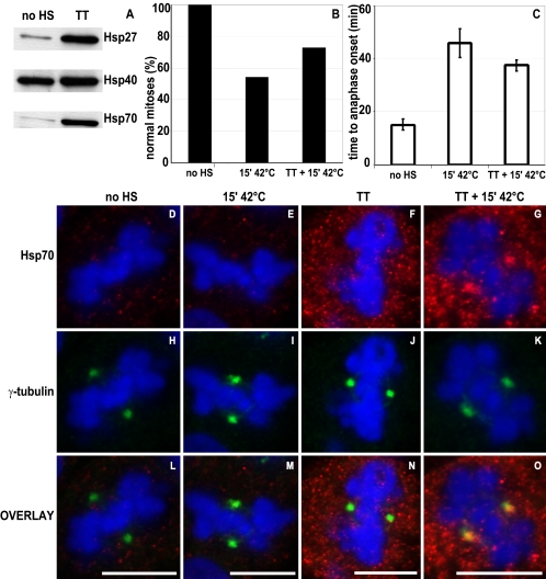
Thermotolerance protects against heat-induced mitotic abnormalities. Cells were left untreated (no HS) or preconditioned to induce thermotolerance (TT). Expression of Hsp27, Hsp40, and Hsp70 was analyzed by Western blotting (A), demonstrating that in TT cells Hsp27, Hsp40, and Hsp70 are induced. Heat shock was given to H2B-GFP–expressing cells (15′ 42°C) or H2B-GFP–expressing TT cells (TT + 15′ 42°C), and mitotic progression was recorded by time-lapse confocal microscopy. Per condition, >20 cells were recorded. The percentage of normal mitoses (B) and mean time ± SD in minutes from early metaphase to anaphase onset (C) are given. After the indicated treatments, cells were methanol/acetone fixed and labeled with monoclonal antibody to Hsp70 (D–G, red) and polyclonal antibody to γ-tubulin (H–K, green). DNA was stained by DAPI (D–O, blue). Overlays of the images are shown in L–O. Bar, 10 μm.

