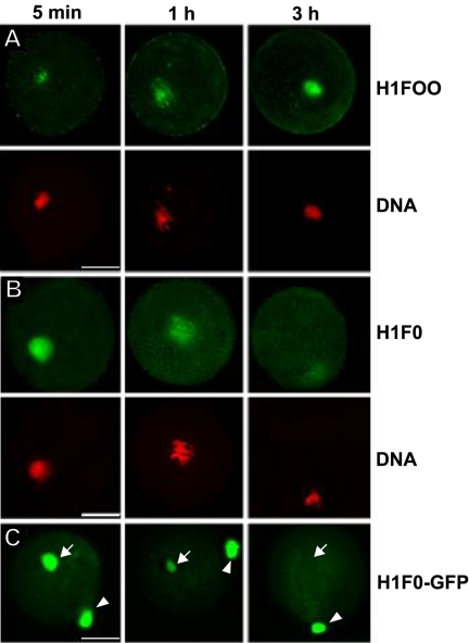
Figure 1.
Exchange of somatic linker histones with oocyte-specific linker histones following transfer of R1 ES cell nuclei. (A-C) Cloned constructs were prepared as previously described (Gao et al., 2003, 2004). R1 ES cell nuclei were injected into enucleated MII-stage oocytes and then cultured as previously described (Gao et al., 2003, 2004) for the indicated time points. (A and B) Exchange of somatic linker histones with H1FOO in chromatin of injected R1 ES cell nuclei. Constructs were fixed and imaged for DNA content, and either oocyte-specific linker histone (A) or somatic linker histone (B) content as described (Gao et al., 2004). (C) H1F0-GFP expressed from a chromosomal locus in a R1 ES cell nucleus is removed from R1 chromatin with a kinetic similar to that of the endogenous somatic H1. To monitor fluorescence loss by bleaching a second nucleus was placed in the perivitelline space next to the ooplasm for comparison. Arrow indicates the injected nucleus; arrow head indicates the nucleus placed in the perivitelline space. Scale bars, 20 μm.

