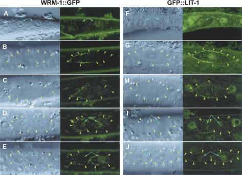
Figure 1.
Localization of WRM-1::GFP and GFP::LIT-1. Anterior is to the left; ventral is to the bottom. Left and right sides of each panel are Nomarski and GFP images of the same area. Cell boundaries are indicated by yellow arrowheads where they are visible (cell boundaries are often not detectable in post-embryonic cells). Positions of the nucleus are indicated by blue arrows. A-E and F-J show the localization of WRM-1::GFP and GFP::LIT-1, respectively. A and F are the T-cell daughters. B-E and G-J are the V5.p cell before division (B,G), in metaphase (C,H), in telophase (D,I), or after the division (E,J).

