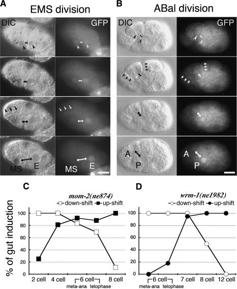
Figure 2.
Cell cycle-dependent asymmetric localization of WRM-1::GFP. (A,B) Nomarski (left columns) and fluorescence micrographs (right columns) of wild-type WRM-1::GFP transgenic animals taken during EMS and ABal cell divisions. (Top panels) White arrowheads indicate the faint, initially similar levels of WRM-1::GFP in early telophase nuclei. Double-headed arrows indicate the nuclei of anterior and posterior sisters. White arrows indicate the cortical localization of WRM-1::GFP. Black arrows indicate the corresponding structures in each Nomarski image. In this and subsequent figures, the bars indicate 10 μm and anterior is to the left. (C,D) Graphic depictions of the temperature-sensitive (ts) periods for gut induction in mom-2(ne874) (C) and wrm-1(ne1982ts) (D). The embryos were either up-shifted (black squares or circles) or down-shifted (white squares or circles) at the indicated cell stage (X-axis), and incubated further to score for gut induction (Y-axis). Shifts were done at two times during the EMS cell cycle: metaphase/anaphase (meta-ana) and telophase (as indicated).

