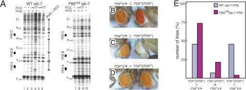
Figure 5.
The PBE is required for PRE silencing in vivo. (A) Binding of PHO/PCC to the iab-7 PRE is abrogated when the PBEs are mutated (for sequences, see Fig. 4A), as revealed by DNaseI footprinting. (B-D) Mutations in the PBE impair PRE-directed PSS. PSS of mini-white by the minimal 260-bp wild-type iab-7 PRE or a mutant, PBE-less iab-7 PRE (PBEmt iab-7 PRE) was compared. Multiple independent lines were established harboring the mini-white transgene under control of either the wild-type or PBEmt iab-7 PRE. For each line, the eye color of homozygotes for the insertion was compared with that of heterozygotes. Representative examples are shown: (B) Homozygotes have a much darker eye color than their heterozygous siblings (P[w+]/P[w+]P[w+]/+), revealing the absence of PSS. (C) Homozygotes have much lighter eyes than heterozygotes ((P[w+]/P[w+][w+]/+), showing strong PSS. (D) The eye color of homozygotes is equal (P[w+]/P[w+] = P[w+]/+) to that of heterozygotes, revealing impaired PSS. (E) The number of lines exhibiting no PSS, impaired PSS, or strong PSS were scored and depicted in a graphical representation with the number of lines displaying a PSS phenotype expressed as percentage of the total number of lines analyzed on the Y-axis and the phenotype on the X-axis. Because the percentages refer to the numbers of independent lines, error bars reflecting the SEM are not applicable. χ2 test of statistical significance for bivariate tabular analysis confirmed the high significance of the difference in PSS frequency between lines harboring either the wild-type or PBEmt iab-7 PREs (χ2 = 12.3, p < 0.01).

