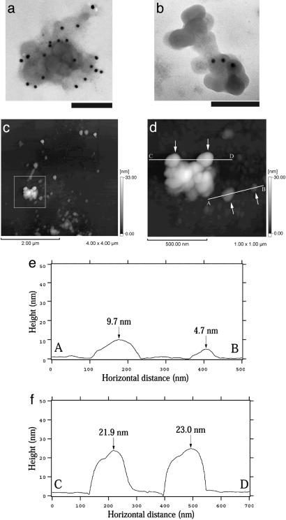
Fig. 3.
Structure of isolated HDQ91 aggregates. (a and b) TEM of uranyl acetate-stained immunogold-labeled HDQ91 aggregates. The size of the gold particles was 10 nm. (Scale bar, 100 nm.) (c) AFM height image of HDQ91 aggregates. (d) Magnified image of the boxed area in c. (e and f) The AFM surface profile along A–B and C–D axes in d, respectively. The arrows in d indicate the globular aggregates, corresponding to the arrows in e and f.

