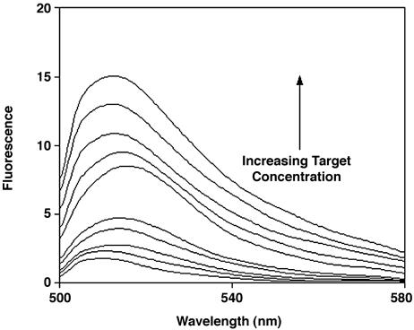
Fig. 5.
A signal transduction mechanism for optical biosensors. Here the target ligand of FynSH3 is detected by folding-linked changes in the emission of a CΔ4-BODIPY conjugate. In the absence of target, the flexibility of the unfolded CΔ4 allows the two tryptophan residues of the protein to quench the fluorescence of the covalently attached BODIPY. Upon ligand binding the tryptophan residues are sequestered, resulting in a large increase in emission. Ligand concentrations are (from bottom up) 0, 0.2, 0.5, 0.8, 1, 2, 5, 8, 10, and 12 mM.

