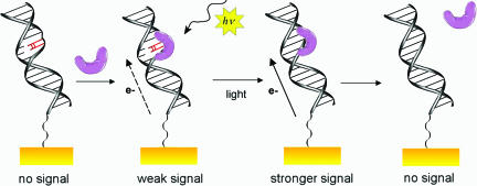
Fig. 7.
Schematic illustration of the electrochemical results. From left to right: In the absence of photolyase, no signal is observed. When the protein (purple) is added, it binds to the T<>T site, and a weak signal is observed. The low signal intensity is due to the disruption of the π-stack, which interrupts communication between the electrode and the protein. Then, upon photoreactivation, the integrity of the π-stack is restored, and the signal grows in intensity. Finally, because the protein has a lower affinity for undamaged DNA, it dissociates, leading to the slow loss of the redox signal.

