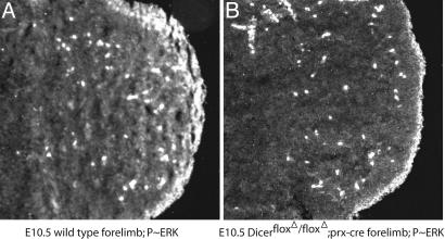
Fig. 7.
FGF signaling is reduced in limb buds lacking Dicer activity. Frontal sections through E10.5 control (A) and Dicerflox/Dicerflox;prx1cre (B) limbs were immunostained with an antibody against phosphorylated ERK/mitogen-activated protein kinase (MAPK), an indicator of active FGF and other RTK signal transduction as described in ref. 20. Phosphorylated ERK/MAPK is detectable at high levels in the mesoderm directly apposed to the apical ectodermal ridge (AER), a potent source of FGF signals, and is present in a gradient extending proximally through the mesoderm from the distal tip (A) (31). In contrast, in limb buds lacking Dicer activity (B), this graded signal is absent, indicating a reduction in FGF/RTK signaling. High levels of phosphorylated ERK/MAPK are still detected beneath the AER, consistent with maintained FGF signaling in the limb ectoderm.

