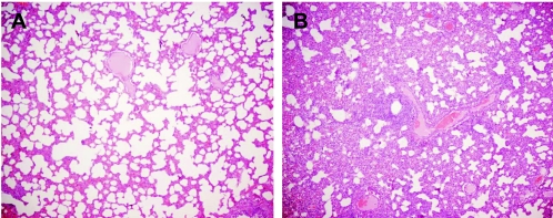
FIG. 3.
Hematoxylin- and eosin-stained sections of lung from an uninoculated pig (A) and a pig infected with A/Vt/1203/04 virus (B). Lung tissues were collected 6 days after inoculation with 3.3 × 10⁶ EID50 of infective virus. In the infected lung tissue (B), the alveoli, interstitial septa, and perivascular spaces are extensively infiltrated by a mixture of inflammatory cells. Magnification, ×4.

