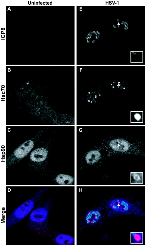
FIG. 1.
Subcellular localization of viral and cellular chaperones in the HSV-1-infected cell. (A to D) Uninfected cells; (E to H) HSV-1-infected cells. Shown are staining profiles for the viral single-stranded DNA binding protein ICP8 (A and E) (green in merged image), for the cellular chaperone Hsc70 (B and F) (red in merged image), and for Hsp90 (shown in blue in panels C and G) (blue in merged image), as well as merged images of triple-labeled cells (D and H). White arrows and insets in panels F through H show the localization pattern of Hsp90 at the VICE foci.

