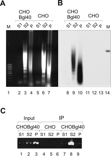
FIG. 6.
The chromatin fractionation of URR-containing plasmid. (A) The nuclei from 107 CHO and CHOBgl40 cells were treated with 30 U of MNase as depicted above (Fig. 5A), and the DNA isolated from each fraction was transferred onto a membrane and hybridized with radiolabeled BPV1 URR-specific probe. Lane 1, 1-kb DNA standard. Lane 14, 100 pg of linearized plasmid Bgl40. (B) After MNase treatment of CHO and CHOBgl40 cells (Fig. 5A), the S1, S2, and P fractions were separated and cross-linked with 1% formaldehyde. The E2/DNA complexes were immunoprecipitated with E2-specific MAb 3E8, and the presence of URR-containing plasmid in the DNA/protein complexes recovered was analyzed by PCR using primers that specifically amplify a region of 7338 to 7654 bp of BPV1 genome. Lanes 1 to 3, the input chromatin from each fraction was tested in parallel with the immunoprecipitated DNA. The image of an agarose gel stained with ethidium bromide is shown.

