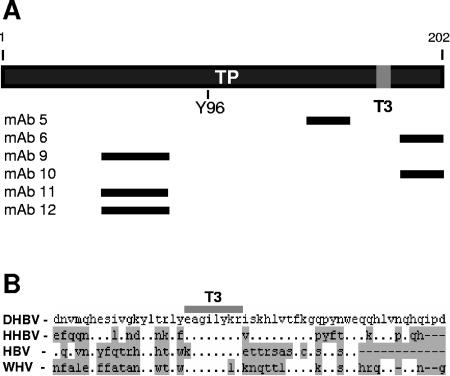
FIG. 3.
Epitope and T3 motif positions within the terminal protein. (A) Regions of the DHBV terminal protein domain containing the epitopes for the MAbs are indicated by black lines. The T3 motif is indicated as a gray block. (B) Alignment of the DHBV, heron hepatitis B virus (HHBV), HBV, and woodchuck hepatitis virus (WHV) terminal domain sequences around the T3 motif (EAGILYKR). Residues differing from those in DHBV are shaded, and gaps are indicated with dashes.

