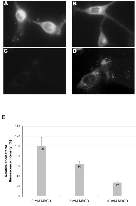
FIG. 3.
Effect of MBCD treatment on the plasma membrane cholesterol level of NIH 3T3 cells. NIH 3T3 cells were treated with (A) 0 mM (mock), (B) 5 mM, or (C and D) 10 mM MBCD at 37°C for 15 min. After this treatment, the cells were fixed and stained with 50 μg/ml filipin for 1 h. Pictures were all taken with an oil-immersion objective (original magnification, 1,000×) using the same camera settings. (A to C) Unprocessed pictures; (D) copy of the picture in C, which has been processed in Adobe Photoshop to visualize the presence of the cells. (E) Quantification of cholesterol extraction. Pictures of mock (0 mM) and MBCD-treated NIH 3T3 cells were analyzed for their fluorescently labeled cholesterol. The cholesterol levels shown are normalized to that of mock-treated cells. At least six randomly taken pictures were measured.

