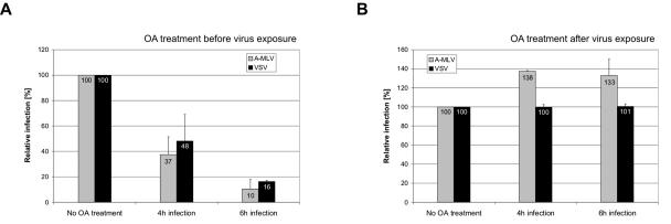
FIG. 6.
Effect of okadaic acid on A-MLV and VSV infection. (A) NIH 3T3 cells were treated with 0.1 μM okadaic acid (OA) for 30 min. Subsequently, the cells were infected with A-MLV or VSV pseudotyped vectors for 4 and 6 h in the presence of okadaic acid. Noninternalized viruses were inactivated using citrate buffer and 48 h after vector addition, infected β-galactosidase-positive cells were counted. The numbers of infected cells are normalized to the mock values. Shown are the means of three (A-MLV) or two (VSV) independent experiments done in duplicate. (B) NIH 3T3 cells were incubated with A-MLV or VSV pseudotyped vectors for 4 or 6 h, washed, and treated with 0.1 μM okadaic acid for 30 min. Subsequently, noninternalized viruses were inactivated using citrate buffer and 48 h after vector addition, infected β-galactosidase-positive cells were counted. The numbers of infected cells are normalized to the mock values. Shown are the means of two independent experiments done in duplicate.

