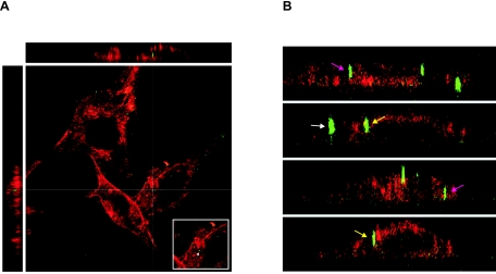
FIG. 7.
Fusion-defective Gag-YFP A-MLV particles can enter NIH 3T3 mouse fibroblasts. (A and B) NIH 3T3 cells were incubated for 6 h with Gag-YFP A-MLV particles (green), which are fusion defective due to an unprocessed envelope protein. The cells were washed with citrate buffer and fixed with paraformaldehyde, and the plasma membrane was stained using rhodamine-labeled concanavalin A (red). The cells were investigated for the presence of intracellular fluorescent viral particles via three-dimensional scanning confocal microscopy. Shown are the YZ (left) and XZ (top) sections with the corresponding XY section. The insert in A shows the virus particle (arrow) at the XY cross-section. (B) XZ and YZ sections. Pink arrows: intracellular particles; yellow arrows: membrane-associated particles; white arrow: extracellular cell-bound particle.

