FIG. 6.
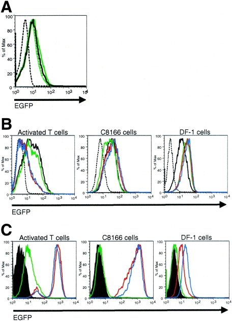
VV uses a unique receptor to bind to activated T cells. (A) T cells were sorted from PBMCs using anti-CD3 microbeads (to >98% purity) and were then activated for 52 h with plate-bound anti-CD3 plus anti-CD28 and anti-CD49d MAbs. Activated T-cells were incubated with rVV-B5R-EGFP or rVV control at an MOI of 50 in the presence or absence of heparin for 1 h at 4°C. Virus binding was assessed by flow cytometry. Dashed black histogram, rVV control; solid black histogram, rVV-B5R-EGFP; solid green histogram, 2 μg/ml heparin, rVV-B5R-EGFP; dashed green histogram, 10 μg/ml heparin, rVV-B5R-EGFP; dotted green histogram, 50 μg/ml heparin, rVV-B5R-EGFP. (B) T cells were sorted from PBMCs using the Pan T Cell Isolation Kit II (to >95% purity) and were then activated with plate-bound anti-CD3 plus anti-CD28 and anti-CD49d for 48 h. Activated T cells, C8166 cells, and DF-1 cells were incubated with preimmune serum or immune sera from mice immunized with activated T cells or monocytes at a 1:10 dilution for 30 min at 25°C and then incubated with rVV-B5R-EGFP or rVV control at an MOI of 50 for 1 h on ice. Virus binding was assessed by flow cytometry. Dashed black histograms, no serum, rVV control; solid black histograms, no serum, rVV-B5R-EGFP; green histograms, preimmune serum, rVV-B5R-EGFP; red histograms, activated T-cell immune serum, rVV-B5R-EGFP; blue histograms, monocyte immune serum, rVV-B5R-EGFP. (C) Cells were incubated with mouse sera as described in the legend to panel B and then stained with a pAb directed against mouse Ig. The ability of the preimmune and immune sera to recognize cellular epitopes was assessed by flow cytometry. Filled histograms, no serum; green histograms, preimmune serum; red histograms, activated T-cell immune serum; blue histograms, monocyte immune serum. These data are representative of three independent experiments.

