
Figure 3.
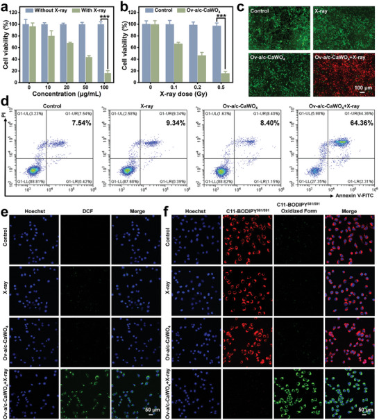
In vitro RDT performance of Ov‐a/c‐CaWO4 NPs. a) Cell viability of 4T1 cells exposed to different concentrations of Ov‐a/c‐CaWO4 NPs for 12 h and then treated with or without X‐ray irradiation (0.5 Gy). n = 3. b) Cell viability of Ov‐a/c‐CaWO4 NPs‐incubated 4T1 cells after exposure to X‐ray irradiation at various doses ([Ov‐a/c‐CaWO4 NPs] = 100 µg mL−1). n = 3. c) Calcein‐AM/PI costaining of 4T1 cells treated with X‐ray irradiation, Ov‐a/c‐CaWO4 NPs, or Ov‐a/c‐CaWO4 NPs plus X‐ray irradiation. d) Flow cytometry analysis of cancer cell apoptosis after diverse treatments. Fluorescence images of 4T1 cells stained with e) DCFH‐DA or f) C11‐BODIPY581/591 after treatment with X‐ray irradiation, Ov‐a/c‐CaWO4 NPs, or Ov‐a/c‐CaWO4 NPs plus X‐ray irradiation (0.5 Gy). Data are presented as mean ± SD. *** p <0.001.