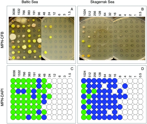
FIG. 3.
Photographs showing MPN-CFB agar plates inoculated with 5 μl from each dilution well/tube. (A) Baltic Sea DCA; (B) Skagerrak DCA. Illustrations of positive growth detected by epifluorescence microscopy after DAPI staining and MPN-DAPI. Blue circles indicate growth detected only by microscopy, representing non-colony-forming bacteria; green circles indicate tubes/wells with growth detected by both microscopy and spot tests on agar plates. These wells/tubes contain colony-forming bacteria and possibly non-colony-forming bacteria. (C) Baltic Sea DCA; (D) Skagerrak DCA. The numbers of bacteria inoculated are indicated in the respective panels.

