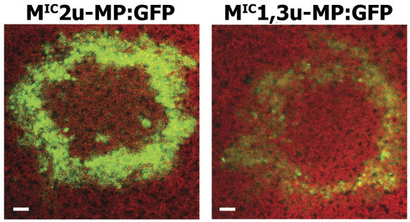
Figure 9.
Cell-to-cell movement of viruses that produce different size VRCs and accumulate different levels of MP:GFP. MIC2u-MP:GFP and MIC1,3u-MP:GFP transcripts were inoculated onto N. benthamiana leaves, and the size and intensity of the fluorescent rings were determined by confocal laser scanning microscopy and example images given. The mutations within the 126-kD protein ORF affected the accumulation, but not the spread, of MP:GFP, representing the location of the virus. Images were taken with the same settings and at the same focal distances. Mean values for ring diameters were 1.42 and 1.39 mm, respectively, for MIC2u-MP:GFP and MIC1,3u-MP:GFP for three replicates (independent rings) per virus (not significant; P > 0.5 by paired t test). The experiment was repeated once with similar results. Bar = 100 μm.

