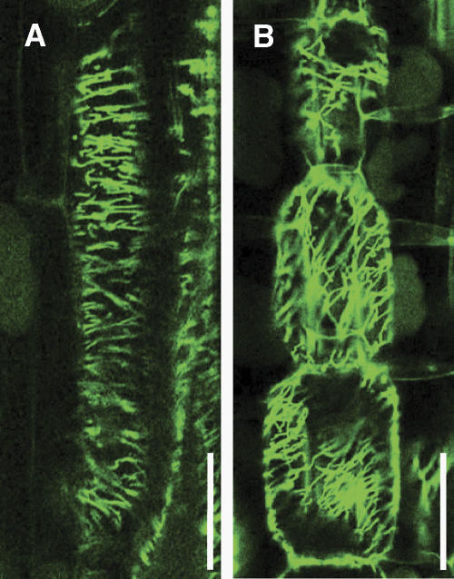
Figure 6.
Arrangement of cortical microtubules in elongating internodal parenchyma cells. Internodal parenchyma cells were subjected to immunofluorescent staining for α-tubulin. The elongating cells in the wild type (A) contain transversely oriented cortical microtubules, whereas dgl1-3 cells (B) contain aberrantly oriented cortical microtubules. Bars = 100 μm.

