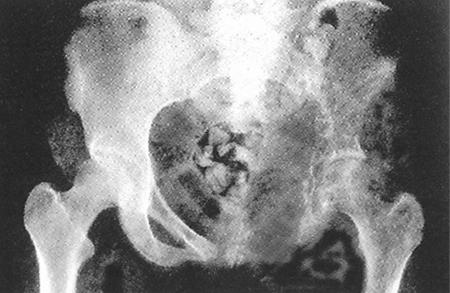
Figure 1.
Antero-posterior radiograph of the pelvis in a patient with Gorham’s disease showing osteolysis involving the left ilium. Note the osseous resorption involving the proximal aspect of the left femur. (Reproduced with permission from Elsevier. In: Peter Bullough, ed. Orthopaedic Pathology. 4th Ed. Philadelphia, PA: Mosby; 2004. Figure 7.58, p. 196. Copyright 2004 Elsevier. All rights reserved).

