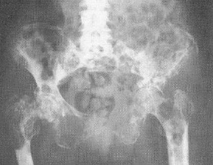
Figure 5.
Plain radiograph of the pelvis of a female patient who has extensive metastatic bone disease secondary to carcinoma of the breast. Note the widespread areas of osteolysis throughout the pelvis and proximal femora. There is also some reactive sclerosis. (Reproduced with permission. In: Peter Renton, ed. Orthopaedic Radiology: Pattern Recognition and Differential Diagnosis. 1st Ed. London, UK: Martin Dunitz Ltd.; 1990. Figure 2.13, p. 86. Copyright 1990 Taylor & Francis. All rights reserved).

