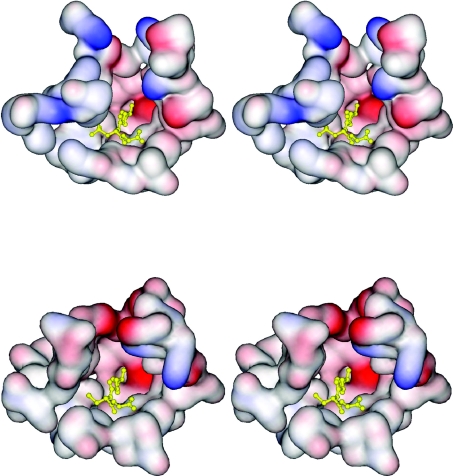
Figure 8. Stereo views of the electrostatic potential surface of the active-site pockets of adGSTD5-5 and adGSTD3-3.
The two tertiary structures were aligned to illustrate the same view of the active site. The top panel shows adGSTD5-5, and the bottom panel shows adGSTD3-3. A yellow ball-and-stick representation shows the GSH in the pocket.

