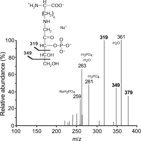
Figure 4. Tandem MS analysis of the phosphorylation product of ribuloselysine by spinach leaf ribulosamine-kinase.
The fragmentation spectrum of the deprotonated, monosodium molecular ion at m/z 379 is shown. The structure of ribuloselysine 3-phosphate and the assignment of the main peaks are also shown.

