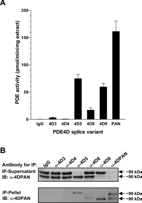
Figure 8. Expression of PDE4D variants in HEK-293 cells.
(A) Cytosolic extracts prepared from HEK-293 cells were subjected to immunoprecipitation with antibodies specifically recognizing PDE4D3, PDE4D4, PDE4D5, PDE4D8 and PDE4D9, as well as the pan-specific PDE4D antibody M3S1 (PAN) and non-immune IgG as a control. The PDE activities of the immunoprecipitation pellets were then determined at 1 μM cAMP. The results represent the means±S.E.M. of 3 experiments. (B) Cytosolic extracts prepared from HEK-293 cells were immunoprecipitated with several splice-form-specific antibodies, as well as the panspecific PDE4D antibody M3S1 (α-4DPAN) and IgG as a control. Both the immunoprecipitation pellet, as well as the depleted immunoprecipitation supernatant, were separated by SDS/PAGE and PDE4D variants were detected by Western blotting using a pan-specific PDE4D antibody.

