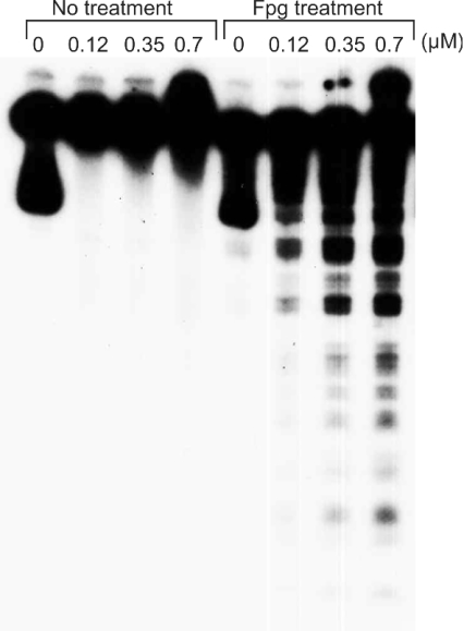
Figure 2. Effect of Fpg treatment on DNA damage induced by histone H1-hydroperoxides in the presence of Cu(I).
Reaction mixtures contained the 32P-5′-end-labelled 337 bp fragment, calf thymus DNA (20 μM in DNA bases), the indicated concentrations of histone H1-hydroperoxides and 20 μM Cu(I) in 200 μl of 10 mM sodium phosphate buffer (pH 7.4) containing 5 μM DTPA. The mixture was incubated at 37 °C for 60 min. The DNA fragments were then treated with Fpg and electrophoresed on an 8% polyacrylamide/8 M urea gel (12 cm×16 cm), and autoradiograms were obtained by exposing an X-ray film to the gel.

